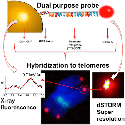
We have just published a neat little paper in ASC Nano, a journal that should be well known to all nano aficionados.
This is a study we performed in collaboration with our colleague Charlie Jeynes from the Centre for Biomedical Modelling and Analysis. Charlie has written a neat summary for nanotechweb.org.
The full title Nanoscale Properties of Human Telomeres Measured with a Dual Purpose X-ray Fluorescence and Super Resolution Microscopy Gold Nanoparticle Probe is a bit of a mouthful. Essentially, we are using a dual gold nanoparticle and organic dye (Alexa 647) probe specific for telomeres. This allows pretty cool combinations of X-ray microscopy (with elemental resolution) and optical super-resolution microscopy. Below we summarise the main findings - for details check out our paper.
Nanoscale Properties of Human Telomeres Measured with a Dual Purpose Probe
 Techniques to analyze human telomeres are imperative in studying the molecular mechanism of aging and related diseases. Two important aspects of telomeres are their length in DNA base pairs (bps) and their biophysical nanometer dimensions. However, there are currently no techniques that can simultaneously measure these quantities in individual cell nuclei. Here, we develop and evaluate a telomere “dual” gold nanoparticle-fluorescent probe simultaneously compatible with both X-ray fluorescence (XRF) and super resolution microscopy. We used silver enhancement to independently visualize the spatial locations of gold nanoparticles inside the nuclei, comparing to a standard QFISH (quantitative fluorescence in situ hybridization) probe, and showed good specificity at ∼90%. For sensitivity, we calculated telomere length based on a DNA/gold binding ratio using XRF and compared to quantitative polymerase chain reaction (qPCR) measurements. The sensitivity was low (∼10%), probably because of steric interference prohibiting the relatively large 10 nm gold nanoparticles access to DNA space. We then measured the biophysical characteristics of individual telomeres using super resolution microscopy. Telomeres that have an average length of ∼10 kbps, have diameters ranging between ∼60–300 nm. Further, we treated cells with a telomere-shortening drug and showed there was a small but significant difference in telomere diameter in drug-treated vs control cells. We discuss our results in relation to the current debate surrounding telomere compaction.