We think that quantitative imaging is the next major goal in advanced imaging. For fluorescence imaging in particular, this is the question how many dye molecules or how many marker molecules are in this image? Ideally, we can relate this information directly to estimate how many proteins, signalling molecules etc are present in our images, with high spatial and temporal resolution. Below we illustrate this with three recent studies.
Quantitative super-resolution imaging of ryanodine receptors
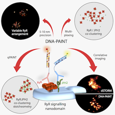 Signalling nanodomains rely on
spatial organisation of proteins to allow controlled intracellular
signalling. Examples include calcium release sites of cardiomyocytes
where ryanodine receptors (RyRs) are clustered with their molecular
partners. Localisation microscopy has been crucial to visualising
these nanodomains, but has been limited by brightness of markers,
restricting the resolution and quantification of individual proteins
clustered within. Harnessing the remarkable localisation precision of
DNA-PAINT (<10 nm), we visualised punctate labelling within these
nanodomains, confirmed as single RyRs. RyR positions within
sub-plasmalemmal nanodomains revealed how they are organised randomly
into irregular clustering patterns leaving significant gaps occupied
by accessory or regulatory proteins. RyR-inhibiting protein
junctophilin-2 appeared highly concentrated adjacent to RyR
channels. Analysing these molecular maps showed significant variations
in the co-clustering stoichiometry between junctophilin-2 and RyR,
even between nearby nanodomains. This is a previously-unseen level of
complexity in RyR arrangement and regulation of calcium signalling,
intrinsically built into the nanodomains.
Signalling nanodomains rely on
spatial organisation of proteins to allow controlled intracellular
signalling. Examples include calcium release sites of cardiomyocytes
where ryanodine receptors (RyRs) are clustered with their molecular
partners. Localisation microscopy has been crucial to visualising
these nanodomains, but has been limited by brightness of markers,
restricting the resolution and quantification of individual proteins
clustered within. Harnessing the remarkable localisation precision of
DNA-PAINT (<10 nm), we visualised punctate labelling within these
nanodomains, confirmed as single RyRs. RyR positions within
sub-plasmalemmal nanodomains revealed how they are organised randomly
into irregular clustering patterns leaving significant gaps occupied
by accessory or regulatory proteins. RyR-inhibiting protein
junctophilin-2 appeared highly concentrated adjacent to RyR
channels. Analysing these molecular maps showed significant variations
in the co-clustering stoichiometry between junctophilin-2 and RyR,
even between nearby nanodomains. This is a previously-unseen level of
complexity in RyR arrangement and regulation of calcium signalling,
intrinsically built into the nanodomains.
The application of refined DNA-PAINT approaches to the molecular characterisation of RyR clusters and JPH2-RyR co-clusters demonstrates the potential for molecular resolution quantitative imaging in complex biological samples. Using all optical methods, it is now possible to obtain data previously thought to be limited to the realm of electron-microscopy. The new data have revealed that the density of RyRs in clusters is lower than expected for dense packing. Clusters follow an irregular assembly pattern compatible with spontaneous self-assembly and sizable gaps in clusters that will affect their calcium signalling. The high resolution of DNA-PAINT data also revealed a previously undetected fraction of JPH2 in molecular proximity of RyRs and presents a novel super-resolution approach to in-situ biochemistry to probe binding candidates in cells and tissues.
For details see
- Isuru D Jayasinghe, Alexander H Clowsley, Ruisheng Lin, T Lutz, Carl Harrison, E Green, David Baddelely, Lorenzo Di Michele, Christian Soeller True molecular scale visualisation of variable clustering properties of ryanodine receptors (2018), Cell Reports.
Quantitative imaging of human neuromuscular junctions
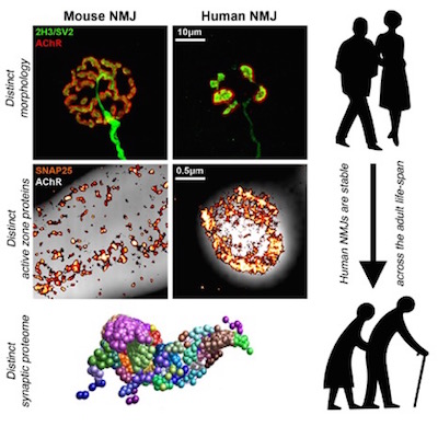 The neuromuscular junction (NMJ) is an experimentally
accessible model synapse routinely studied in animal models to explore
fundamental aspects of synaptic form and function. Synapses play
fundamental roles in the form and function of the nervous system both
in health and during disease. Despite numerous important breakthroughs
in our understanding of the cellular and molecular composition of
synapses in animal models, we know surprisingly little about the
equivalent make up of synapses in humans. Current studies of synaptic
connectivity at the cellular and molecular level have therefore relied
heavily on ‘model’ organisms, both vertebrate and invertebrate,
working on the tacit assumption that the biological principles
uncovered can ultimately be applied to humans.
The neuromuscular junction (NMJ) is an experimentally
accessible model synapse routinely studied in animal models to explore
fundamental aspects of synaptic form and function. Synapses play
fundamental roles in the form and function of the nervous system both
in health and during disease. Despite numerous important breakthroughs
in our understanding of the cellular and molecular composition of
synapses in animal models, we know surprisingly little about the
equivalent make up of synapses in humans. Current studies of synaptic
connectivity at the cellular and molecular level have therefore relied
heavily on ‘model’ organisms, both vertebrate and invertebrate,
working on the tacit assumption that the biological principles
uncovered can ultimately be applied to humans.
It is surprising how little is currently known about NMJs in humans. Here, we combined morphological techniques, super-resolution imaging and proteomic profiling to reveal the detailed cellular and molecular architecture of the human NMJ. Human NMJs were significantly smaller, less complex and more fragmented than mouse NMJs. In contrast to data from animal studies, human NMJs were also remarkably stable across the entire lifespan. Super-resolution imaging and proteomic profiling revealed distinctive active zone architecture and expression of core synaptic proteins and pathways at the human NMJ. Taken together, these findings reveal unexpected human-specific features of the NMJ that distinguish them from comparable synapses in other mammalian species.
For details see
- Ross A Jones, Carl Harrison, Samantha L Eaton, Maica Llavero Hurtado, Laura C Graham, Leena Alkhammash, Oladayo A Oladiran, Andy Gale, Douglas J Lamont, Hamish Simpson, Martin W Simmen, Christian Soeller, Thomas M Wishart, Thomas H Gillingwater Cellular and Molecular Anatomy of the Human Neuromuscular Junction (2017), Cell Reports, 21:2348-2356.
Quantitative imaging of human telomeres with optical super-resolution and x-ray fluorescence
 Techniques to analyze human telomeres are imperative in
studying the molecular mechanism of aging and related diseases. Two
important aspects of telomeres are their length in DNA base pairs
(bps) and their biophysical nanometer dimensions. However, there are
currently no techniques that can simultaneously measure these
quantities in individual cell nuclei. Here, we develop and evaluate a
telomere “dual” gold nanoparticle-fluorescent probe simultaneously
compatible with both X-ray fluorescence (XRF) and super resolution
microscopy. We used silver enhancement to independently visualize the
spatial locations of gold nanoparticles inside the nuclei, comparing
to a standard QFISH (quantitative fluorescence in situ hybridization)
probe, and showed good specificity at ∼90%. For sensitivity, we
calculated telomere length based on a DNA/gold binding ratio using XRF
and compared to quantitative polymerase chain reaction (qPCR)
measurements. The sensitivity was low (∼10%), probably because of
steric interference prohibiting the relatively large 10 nm gold
nanoparticles access to DNA space. We then measured the biophysical
characteristics of individual telomeres using super resolution
microscopy. Telomeres that have an average length of ∼10 kbps, have
diameters ranging between ∼60–300 nm. Further, we treated cells with a
telomere-shortening drug and showed there was a small but significant
difference in telomere diameter in drug-treated vs control cells. We
discuss our results in relation to the current debate surrounding
telomere compaction.
Techniques to analyze human telomeres are imperative in
studying the molecular mechanism of aging and related diseases. Two
important aspects of telomeres are their length in DNA base pairs
(bps) and their biophysical nanometer dimensions. However, there are
currently no techniques that can simultaneously measure these
quantities in individual cell nuclei. Here, we develop and evaluate a
telomere “dual” gold nanoparticle-fluorescent probe simultaneously
compatible with both X-ray fluorescence (XRF) and super resolution
microscopy. We used silver enhancement to independently visualize the
spatial locations of gold nanoparticles inside the nuclei, comparing
to a standard QFISH (quantitative fluorescence in situ hybridization)
probe, and showed good specificity at ∼90%. For sensitivity, we
calculated telomere length based on a DNA/gold binding ratio using XRF
and compared to quantitative polymerase chain reaction (qPCR)
measurements. The sensitivity was low (∼10%), probably because of
steric interference prohibiting the relatively large 10 nm gold
nanoparticles access to DNA space. We then measured the biophysical
characteristics of individual telomeres using super resolution
microscopy. Telomeres that have an average length of ∼10 kbps, have
diameters ranging between ∼60–300 nm. Further, we treated cells with a
telomere-shortening drug and showed there was a small but significant
difference in telomere diameter in drug-treated vs control cells. We
discuss our results in relation to the current debate surrounding
telomere compaction.
For details see
- J Charles G Jeynes, Kalotina Geraki, Christopher Jeynes, Mi Zhaohong, Andrew A Bettiol, Eva Latorre, Lorna Wendy Harries, Christian Soeller Nanoscale Properties of Human Telomeres Measured with a Dual Purpose X-Ray Fluorescence and Super Resolution Microscopy Gold Nanoparticle Probe (2017), ACS nano, acsnano.7b07064.